Quotation
Host Cell Protein (HCP), as the most significant process-related impurity in biopharmaceutical production, its residue level is directly related to the safety and efficacy of the product. For a long time, the industry has generally used the ELISA method to quantitatively measure the total amount of HCP, and has taken "ng/mg", or "ppm", as the single quality control index[1] [2]. However, as the understanding of the biological functions of HCP deepens, more and more evidence indicates that the risks of HCP do not equate to its total amount - trace amounts of protease may degrade the product, proteins with immune adjuvant activity may cause adverse reactions, while some high-abundance structural proteins pose almost no safety risks.
This perception is driving the HCP quality control concept to transform from "total quantity monitoring" to "risk classification". In 2021, the BPDG (Biopharmaceutical Process Development Group) consensus document, jointly published by 26 biopharmaceutical enterprises, systematically proposed for the first time the four major classification frameworks of high-risk HCPS[3]; The USP chapter <1132.1>, which came into effect in May 2025, incorporates mass spectrometry technology into the regulatory framework for HCP detection, providing a methodological basis for risk identification at the single-protein level[4]。
This series of tweets will be divided into four chapters, systematically sorting out the upgrade path of the HCP quality control system:
Chapter One explores the evolution of HCP risk cognition, from the limitations of traditional total quantity monitoring to the four major risk classification frameworks of BPDG, as well as the characteristic differences of high-risk HCPS in different expression systems.
Chapter Two conducts an in-depth analysis of the six root causes of the differences in ELISA method reports, reveals the influence mechanism of the mismatch of calibrator composition through quantitative models, and proposes the principle of process matching and the selection strategy of single-cell line calibrators.
Chapter Three elaborates on the full life cycle selection strategy of HCP detection methods, including the method selection at different stages of product development, the six-step decision-making framework for candidate method screening, as well as the risk assessment and regulatory path for method changes.
Chapter Four explores the collaborative application of orthogonal techniques and new regulatory developments, including the technological evolution of antibody coverage assessment, the role upgrade of LC-MS in HCP detection and USP <1132.1>, as well as targeted detection strategies for high-risk HCP.
As the first post in a series of tweets, this article will first focus on the evolution of HCP risk awareness.
The Evolution of HCP Risk PerceptionFrom "Total Quantity" to "Individual"
PART 01
The limitations of traditional total quantity monitoring
The core logic of traditional HCP quality control is: assuming that the risks of all HCPS are equal, safety can be guaranteed as long as the total amount is controlled below the threshold. This logic faces three fundamental challenges in practice:
01
The high heterogeneity of HCP composition
A typical example is that thousands of different HCPS can be identified in CHO cell harvest fluid, with their abundance ranging from several orders of magnitude[2]. During the purification process, the removal efficiency of different HCPS varies greatly - ion-exchange chromatography may selectively enrich specific HCPS with charge properties similar to those of the product, while affinity chromatography may allow certain chaperone proteins to "free-ride" for co-purification[2] [3]。
The composition of the residual HCP in the final stock solution was significantly different from that in the harvest solution: the downstream purification conducted a strong selective screening of HCP. The high-abundance proteins that were easy to remove were largely eliminated, while the co-purified proteins that were similar in physicochemical properties to the product and difficult to remove were relatively enriched. Commercial reagent kits generally use upstream harvest solutions to prepare calibrators, which is a common standard practice in the industry. When the process matching degree is high, it can meet the requirements of process optimization and trend monitoring. However, when the cell lines, culture processes or purification procedures of the calibrators differ significantly from those of the actual samples, the mismatch in protein composition and antibody reactivity between the two can introduce systematic biases, causing the reported values to deviate from the true residual levels.
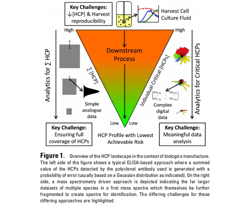
Figure 1 Overview of HCP analysis in biomanufacturingThe differences in the analysis methods of total HCP and key HCP in upstream/downstream processes
02
The risk levels of different HCPS vary greatly
A typical case is phospholipase B-like protein 2 (PLBL2) : In some monoclonal antibody products, the residual amount of PLBL2 accounts for less than 5% of the total HCP. However, due to its lipase activity and co-purification with the product, it is classified as a high-risk HCP by BPDG and has received significant attention from the industry and regulatory authorities[3]. On the contrary, some high-abundance cytoskeletal proteins (such as actin and tubulin), although accounting for a high proportion of the total HCP, usually have no immunogenicity or enzymatic activity, and the actual risk is limited[2][6]。
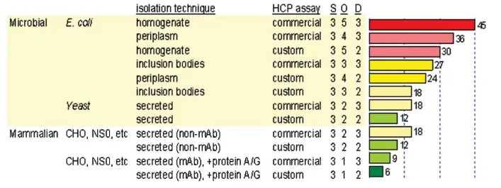
Figure 2 Risk Analysis (FMEA) of HCP control: Severity (S)× incidence (O)× detectability (D) score of different expression systems and purification techniques.
The higher the risk score (red), the greater the residual risk of HCP; the lower the score (green), the more controllable the risk (Wang et al., 2009[2])
03
The total ELISA data cannot reflect the risk of single proteins
The ELISA method provides a summary value and cannot inform the user of which specific proteins are included, what proportion they account for, or whether there are known high-risk factors[1] [2]. When the total HCP of the two products is both 10 ng/mg, one is mainly composed of PLBL2 and the other is mainly composed of inactive structural proteins. Their safety risks are completely different, but ELISA cannot distinguish them.
PART 02
The four major risk classification frameworks of BPDG
In 2021, Jones et al. published a multi-enterprise collaborative study in Biotechnology and Bioengineering, systematically sorting out 25 high-risk HCPS in the CHO expression system for the first time and classifying them into four major categories according to the risk mechanism:
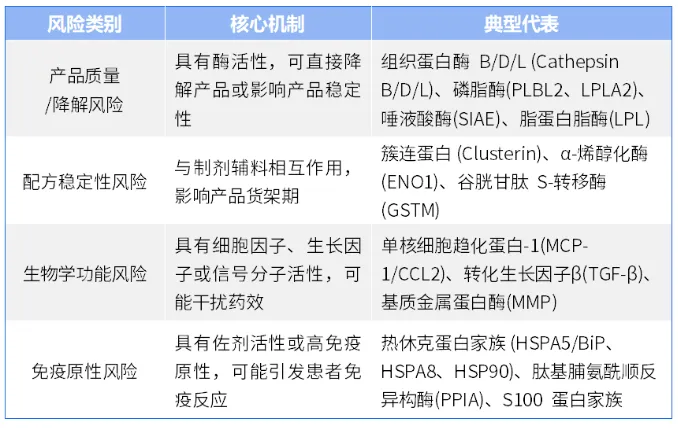
In addition, the study specifically pointed out the fifth type of risk -Co-purified/difficult-to-remove HCP. These proteins themselves may not have direct toxicity or immunogenicity, but due to their similar physicochemical properties (charge, hydrophobicity, molecular weight) to the product molecules, they are difficult to remove through conventional chromatography steps and often become the main contributors to HCP residues in the stock solution[2]. Typical representatives include protein disulfide isomerase (P4HB), voltage-dependent anion channel protein (VDAC), and various ribosomal proteins.
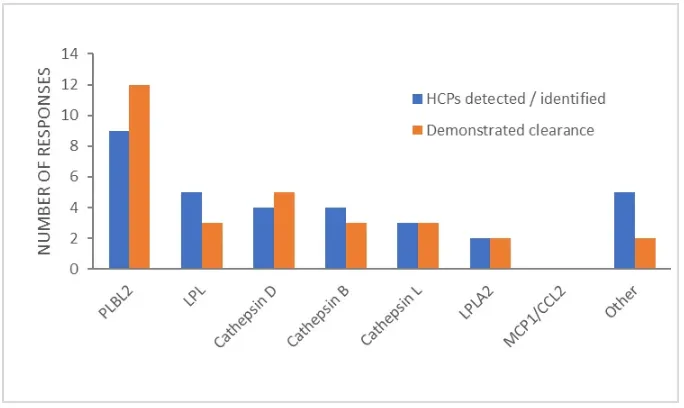
Figure 3 Survey Results of high-risk HCP industries in BPDG:
The quantity of each high-risk HCP detected/identified in the final product (blue) compared with the quantity that has been proven to be cleared (orange).
PLBL2 is the most frequently detected high-risk HCP and also the subject with the most clearance studies (Jones et al., 2021).
PART 03
Differences in high-risk HCPS among different expression systems
The spectra of high-risk HCPS have strong expression system specificity. Directly applying the high-risk list of the CHO system to E. coli, HEK293 or other expression systems may lead to the omission of key risks. The following is a comparison of the high-risk HCP characteristics of the main expression system:
01
E. coli expression system
Unlike mammalian cells, the most prominent risk in E. coli HCP is immunogenicity. Flagellin (FliC), as a potent ligand of TLR5, may trigger a strong innate immune response even with trace residues[6]; Heat shock proteins (DnaK, GroEL, IbpB) and outer membrane proteins (OmpA, OmpF) also have significant adjuvant effects[6]. In addition, the proteases specific to E. coli (Lon, DegP, Ptr) and amidase (AmiD) may cause degradation of recombinant protein products[1] [7]. It is worth noting that there are significant differences among different strains: K-12-derived strains such as W3110 do not express the flagellar system due to the absence of FliA (σ28), while the BL21 strain has defects in Lon and OmpT proteases.
02
The HEK293 expression system
In gene therapy and vaccine products, the high-risk HCP of HEK293 presents a unique spectrum. LGALS3BP (G3BP), as an endogenous ligand of TLR4, has a tendency to co-purify with the AAV capsid and is regarded as a highly risky factor[6]; Molecular chaperone networks (HSPA1B, HSP90AB1, ST13) dominate the composition of residual HCPS in AAV production. Apoptosis-related caspase-3/8 was significantly upregulated during the production of viral vectors. Nucleic acid binding proteins (NCL, NPM1, YB-1) may be co-purified with the AAV genome[2] [6]。
03
MDCK expression system
As the main production cell line of influenza vaccines, the high-risk HCP of MDCK has canine specificity. GSTM4 shows abnormally high abundance expression in MDCK. ATP1A1 (sodium-potassium ATpase α1 subunit) is the membrane protein with the highest abundance. CYP4A47 is a cytochrome P450 enzyme that is highly expressed specifically in dogs[2]. In addition, trypsin is usually added during the production of influenza vaccines for virus lysis. Although this exogenous protease (typically from cattle or pigs) is not canine HCP, it needs to be monitored separately as a process-related impurity.
04
SF9 Insect cell expression system
The HCP risk of the baculovirus-insect cell expression system is unique. Sf-cathepsin L is currently the only SF9 endogenous protease that has been clearly identified. Sf-caspase-1 (P89116) is the main apoptotic effect caspase and is released in large quantities in the later stage of baculovirus infection[2] [6]. Since the homology between insect proteins and human proteins is usually only 40% to 60%, immunogenicity is the most prominent risk dimension of the SF9 system[6]. Furthermore, the proteins encoded by baculovirus itself - including the envelope glycoprotein GP64 (P17501), viral cathepsin V-CATH (P25783), and chitinase CHIA (P41684) - need to be included in the risk assessment as key process impurities[2]. Another special challenge of the SF9 system is that the cell viability at harvest is usually only 20% to 50%, and the intracellular proteins released by a large amount of cell lysis lead to an extremely high HCP load[2]。
The high-risk HCP spectra in this section are comprehensively compiled from references [2], [3], [6] and proteomic studies of various expression systems
Summary of this chapter
From "total quantity monitoring" to "risk classification", it is not only an upgrade of HCP's quality control concept, but also a deepening of the understanding of the safety risks of biopharmaceutical products. Traditional total quantity monitoring assumes that all HCPS have equal risks, while the risk classification concept recognizes that the composition of HCPS is highly heterogeneous, and the risk levels of different HCPS vary greatly. ELISA total quantity data cannot reflect the risk of individual proteins.
The four major risk classification frameworks of BPDG provide us with a systematic tool for identifying high-risk HCPS. However, the differences in the spectra of high-risk HCPS in different expression systems remind us that there is no "universal" HCP detection method. Targeted selection must be made based on the specific expression system and process characteristics of the product.
Next part Preview
In the next article, we will delve into the six major sources of ELISA method reporting differences and analyze the impact of calibrator composition mismatch on quantitative results through quantitative models. We will also detail the principle of process matching and the selection strategy of single-cell line calibrators. Please stay tuned.
Product Link

References
[1] USP General Chapter <1132> Residual Host Cell Protein Measurement in Biopharmaceuticals.
[2] Wang X, Hunter AK, Mozier NM. Host cell proteins in biologics development. Biotechnol Bioeng. 2009;103(3):446-458.
[3] Jones et al. "High-risk" host cell proteins (HCPs): A multi-company collaborative view. Biotechnol Bioeng. 2021;118(7):2561-2572.
[4] USP General Chapter <1132.1> Residual Host Cell Protein Measurement in Biopharmaceuticals by LC-MS. USP-NF 2025.
[5] Bracewell DG, Francis R, Smales CM. The future of host cell protein (HCP) identification during process development and manufacturing linked to a risk-based management for their control. Biotechnol Bioeng. 2015;112(9):1727-1737. DOI:10.1002/bit.25627.
[6] Haltaufderhyde K, et al. Immunoinformatic Risk Assessment of Host Cell Proteins.
0756-8699969
Address: No. 88, Shuian 1st Road, Xiangzhou District, Zhuhai City, Guangdong Province
Email: marketing@biori.com
Wechat official account |
Product consultation |
Video Account |